13/06/2018

First Commercially Available Active Human and Mouse ASYN Proteins from StressMarq


Alpha-synuclein is a 140 amino acid protein which in humans, is encoded by the SNCA gene. It is expressed predominantly in the brain and particularly in presynaptic nerve terminals.

Due to its structural flexibility, alpha-synuclein can adopt several conformations and depending on the environment and the binding partners, exists as a dynamic balance between monomeric unfolded and an amphipathic alpha-helix (membrane binding) states.

In healthy brains, quality control systems ensure the correct assembly of alpha-synucleins and intracellular alpha-synuclein homeostasis is controlled via the ubiquitin-proteasome system and the lysosomal autophagy system, with the latter being involved in clearing oligomer assemblies. Other synucleins are also able to inhibit and control the oligomerisation of alpha-synuclein.

Failure in these systems, oxidative stress, pH changes are, to name a few, examples of triggers that can lead to the overproduction and accumulation of alpha-synuclein. In addition, post-translational modifications of alpha-synuclein can lead to a change in conformation resulting in the alpha-synuclein proteins being more susceptible to aggregation. In Lewy bodies, it has been shown that phosphorylated Ser129 alpha-synuclein is the most abundant form of alpha-synuclein present in these aggregates.

In pathological conditions such as Parkinson’s disease, soluble alpha-synuclein monomers associate to form oligomers that will combine further to generate protofibrils that subsequently aggregate to form large and insoluble aggregates, the main component of Lewy body inclusions.[1] These neurotoxic aggregates will cause the progressive degeneration of dopaminergic neurons in the substantia nigra pars compacta, causing the motor symptoms in PD. [2]

StressMarq has recently released the first commercially available active Human and Mouse Alpha Synuclein Monomers and Pre-formed Fibrils:

- Active Human Recombinant Alpha Synuclein Protein Monomer (Cat No. SPR-321)
- Active Human Recombinant Alpha Synuclein Pre-Formed Fibrils (Cat No. SPR-322).
- Active Mouse Recombinant Alpha Synuclein Protein Monomer (Cat No. SPR-323)
- Active Human Recombinant Alpha Synuclein Pre-Formed Fibrils (Cat No. SPR-324).

The active alpha synuclein pre-formed fibrils (Cat No. SPR-322) in presence of the active alpha-syn monomer (SPR-321) has been shown to induce Lewy body inclusion formation in neuronal cell culture, a characteristic of Parkinson’s Disease.

This was demonstrated using T-thioflavin, a fluorescent dye that binds to beta sheet-rich structures, such as those in α-Syn aggregates and which upon binding, experiences a red-shift in its emission spectrum and increased fluorescence intensity. As seen in the images below, it was shown that 10 nM of active α-Syn aggregate (SPR-322) combined with 100 µm of active α-Syn monomer (SPR-321) could induce aggregation, as compared to active α-Syn aggregate (SPR-322) and active α-Syn monomer (SPR-321) alone.


A similar experiment was also performed to show the activity of the active mouse pre-formed fibrils and monomers (SPR 323 and SPR324).


In addition, StressMarq has also launched control alpha synuclein protein monomers (Cat No. SPR-316) and control alpha synuclein protein aggregates (Cat No. SPR-317) which are inactive as shown in the image below.


These active alpha synuclein proteins and the related alpha synuclein antibodies below can be purchased in the UK and Ireland via Newmarket Scientific, the distributor of StressMarq.

StressMarq alpha synuclein antibodies: Cat. No.
Alpha synuclein, clone 3C11 SMC-530
Alpha synuclein, clone 10H7 SMC-531
Alpha synuclein, clone 3F8 SMC-532
Alpha synuclein, clone 4F1 SMC-533
Alpha synuclein pSer129 SPC-742

References:
[1] Alpha-Synuclein: From Early Synaptic Dysfunction to Neurodegeneration, Ghiglieri V. et al, Front Neurol. 2018; 9 : 295

[2] Linking Neuroinflammation and Neurodegeneration in Parkinson’s Disease, Gelders G. et al, J Immunol Res., 2018: 4784268. doi: 10.1155/2018/4784268. eCollection 2018.

Written by Magalie Dale
If you like my post why not connect to me on LinkedIn.

22/05/2018

Migraine: The culprits behind the headache?

Migraine is not just a simple headache. Very often it is accompanied by various symptoms such as nausea, sensitivity to light, noise and visual disturbances, it is characterised by a unilateral throbbing pain sensation that can last for up to 72 hours and is accentuated during physical exercise. 1 in 7 people suffers from migraine and although it is not correlated with age or social background, women tend to suffer twice as much than men. In the UK alone, it is estimated that migraine costs £2 billion a year, hence extensive research is being carried out to develop effective treatments.[1]

Headaches in Migraine – The Crucial Role of CGRP
The headache phase in the migraine occurs when the dilated blood vessels mechanically activate the perivascular trigeminal sensory nerve fibres, the principle sensory nerve in the head. This triggers a pain response with several neurotransmitters, such as substance P and CGRP (calcitonin gene–related peptide), being released to transmit the nociceptive signals from the trigeminal sensory afferents to second-order neurons. CGRP, a potent vasodilator, will aggravate the dilation of cranial blood vessels, cause mast-cell degranulation and initiate neurogenic inflammation within the meninges. As a result, prolonged activation of the meningeal trigeminal nerves, vessels and mast cells causes sensitisation of secondary and tertiary-order neurons which could explain symptoms associated with migraine such as allodynia (skin sensitivity) and photosensitivity.[2]

The role played by CGRP in migraine headaches is widely accepted. Indeed, it has been observed that during a migraine attack, the levels of CGRP in blood and saliva were increased.[3] It was also shown that administrating CGRP to patients prone to migraines resulted in inducing migraine-like headaches.[4]

The Controversial Role of TRPV1 in Migraine Pathology
More recently, it has been shown that the cation channel TRPV1, a nonselective cation channel that is activated by stimuli such as high temperature and capsaicin could also be involved in migraine pathophysiology. Colocalised with CGPR in trigeminal ganglion neurons, TRPV1 when activated promotes the release of CGPR.

It is also known that patients suffering from chronic migraine show elevated levels of nerve growth factor (NGF) in the cerebral spinal fluid and it has been suggested that a NGF-dependent mechanism could lead to the insertion of TRPV1 into the plasma membrane hence increasing the number of TRPV1 channels available for activation on the nociceptor surface membrane. In addition, several studies have also demonstrated that through different pathways, prostaglandins, ATP, BK and possibly NGF reduce the TRPV1 activation threshold by phosphorylating TRPV1 on S502 and S800 and hence causing sensitisation.[5]

Based on this, TRPV1 antagonists seemed to be attractive targets for the treatment of migraine and SB-705498, a TRPV1 antagonist showed promising results in cats. However, in the case of humans, the clinical trial was terminated early due to a lack of efficacy in treating acute migraine.[6] More recently, two TRPV1 receptor antagonists have been shown to be effective in two different experimental models of migraine suggesting that further clinical trials using different TRPV1 antagonists should be performed to better understand the role played by TRPV1 in migraine pathology.[6]

Working with TRPV1? Have a look at our products:

Biosensis Antibodies to TRPV1 Cat. No.
Rabbit antibody to human capsaicin receptor (531-541): whole serum R-053-100
Rabbit antibody to human capsaicin receptor (608-621): whole serum R-076-100
Mouse monoclonal antibody to rat capsaicin receptor (VR1, TRPV1, 819-838), [Clone BS397]: IgG M-1714-100
StressMarq Small Molecules Cat. No.
Capsaicin (TRPV1 opener) SIH-322
BCTC (TRPV1 blocker) SIH-307
SB-366791 (TRPV1 blocker) SIH-321

Current and Emerging Treatments


Current migraine treatments include the use of nonsteroidal anti-inflammatory drugs (NSAID) such as aspirin, ibuprofen, naproxen and didofenac potassium. When the pain is moderate to severe, triptans (highly selective serotonin 5-HT1B and 5-HT1D receptor agonists) are considered to be the first line of treatment. Indeed, serotonin vasoconstricts the nerve endings and blood vessels and consequently affects nociceptive pain. [7]

However, because of the presence of the 5-HT1B receptors on blood vessels and the vascular risks associated with the use of these drugs, ditans, a novel class of chemicals selectively targeting the 5-HT1F receptors expressed in the trigeminal nerve pathway but lacking the vasoconstrictive properties, are being developed. Currently, lasmiditan is in phase III clinical trials in the US. [8]

As seen previously, CGRP plays an important role in the pathogenesis of migraine and as a result, several small molecules, CGRP receptor antagonists called gepants have been developed. In particular, telcagepant and MK-3207 were shown to be effective treatments for acute migraine, but were terminated due to their liver toxicity.[2] Other drugs based on monoclonal antibodies are currently being developed. These new treatments target either CGRP or the CGRP receptors and currently show promising results in human clinical trials. Aimovig (erenumab) developeed by Amgen and Novartis has recently been FDA-approved as a preventive migraine treatment by blocking the activity of CGRP receptors.[9]

Migraine Research: ImmunoStar Antibodies can help
ImmunoStar has developed an excellent range of antibodies for neuroscience research. These antibodies are put through extensive testing before release to ensure they are both high quality and high titer. This provides excellent reliability and lot-to-lot consistency. They have also been specifically tested for use in immunohistochemistry. They are currently available to purchase through Newmarket Scientific.

Migraine related articles using antibodies from the ImmunoStar range can be found below:

References ImmunoStar antibodies
Neural mechanism for hypothalamic-mediated autonomic responses to light during migraine,
Noseda R et al, PNAS, 2017, 114 (28): E5683-E5692
Tyrosine hydroxylase, oxytocin, vasopressin 1
Hypothalamic and basal ganglia projections to the posterior thalamus: Possible role in modulation of migraine headache and photophobia;
Kagan R et al, Neuroscience 2013, 248, 359-368
Tyrosine hydroxylase, cholecystokinin octapeptide (CCK8)
Neurochemical Pathways That Converge on Thalamic Trigeminovascular Neurons: Potential Substrate for Modulation of Migraine by Sleep, Food Intake, Stress and Anxiety,
Noesda R et al, Plos one 2014, 9 (8): e103929
Tyrosine hydroxylase
Dopamine β-Hydroxylase Statins Decrease Expression of the Proinflammatory Neuropeptides Calcitonin Gene-Related Peptide and Substance P in Sensory Neurons,
Bucelli RC et al, J. Pharmacol Exp Ther, 2008, 324 (3): 1172-1180 
Substance P

References:
[2] Migraine, Dodick DW, The Lancet, 2018, 391 (10127), 1315-1330
[3] Vasoactive peptide release in the extracerebral circulation of humans during migraine headache. Goadsby PJ et al, Ann Neurol. 1990;28:183–7.
[4] Elevated saliva calcitonin gene-related peptide levels during acute migraine predict therapeutic response to rizatriptan. Cady RK et al. Headache. 2009;49:1258–66.
[5] TRPV1 in migraine pathophysiology, Meents JE et al, Trends in Molecular Medicine, 2010, 16 (4):153-159
[6] Two TRPV1 receptor antagonists are effective in two different experimental models of migraine, Meents JE et al, The Journal of Headache and Pain, 2015, 16:57
[7] CGRP and serotonin in migraine, Aggarwal M et al, Annal od Neurosciences, 2012 19 (2): 88-94 https://www.researchgate.net/publication/265558756_Serotonin_and_CGRP_in_migraine
[8] Migraine Therapy: Current Approaches and New Horizons,  Goadsby PJ et al, Neurotherapeutics, 2018, 15 (2) :271-273
[9] https://www.genengnews.com/gen-news-highlights/amgen-novartis-set-to-launch-migraine-drug-aimovig-next-week-after-fda-approval/81255834 Accessed on 21/05/2018


Written by Magalie Dale
If you like my post why not connect to me on LinkedIn.

09/05/2018

Amyloid beta plaque staining

Alzheimer's disease is characterised by a gradual decline of the cognitive functions and it is accepted that one of its hallmarks is the accumulation of insoluble proteinaceous deposits called amyloid fibrils. Originally thought to be carbohydrate in nature as they could be stained in tissues with iodine, they were called amyloids as iodine was a well-known stain for polysaccharides such as amylose. It was later discovered they were actually constituted of proteins fibres (mostly amyloid beta 1-40 and amyloid beta 1-42), but for historic reasons, the name was conserved.
Histologic dyes such as Congo Red or Thioflavin S or T show affinity for the amyloid plaques. Although they are commonly used for amyloid plaques detection, they come with several limitations, firstly in their spectral properties, with both having a broad range of excitation frequencies leading to a "bleed through" when illuminated with other filters and secondly by requiring harsh chemicals making it less compatible with subsequent use of antibodies.
Amylo-Glo RTD (Cat. No. TR-300-AG) is a new histological marker developed by Biosensis (Thebarton, South Australia) that shows clear superiority for visualising amyloid plaques over these conventional markers (Schmued et al., 2012).
  1. Amylo-Glo shows the same affinity as conventional probes (Congo red, ThioflavinS and Pan abeta 40) and is suitable to use with fresh, frozen, and formalin-fixed immunohistochemistry or cytochemistry.
  2.  Amylo-Glo has a unique emission/excitation profile. Only excitable with UV light and emits a bright blue/yellow colour.
  3.  Amylo-Glo only uses mild conditions making it ideal for subsequent immunohistochemistry labelling.
  4.  Its chemical and spectral properties allow for multi-labelling studies. Its specific blue fluorescence contrasts effectively with red and green immunofluorescence labelling.
  5. Amylo-Glo is perfect for low magnification studies. Indeed, its unusual brightness (5-6 times brighter than conventional probes) allows to generate strong signals at extremely low magnification (x2).

This triple exposure allows for the simultaneous localization of Amylo-Glo® positive amyloid plaques (blue), GFAP positive hypertrophied astrocytes (green) and activated microglia (red) in the hippocampus of the AD/Tg mouse. Combined UV, blue and green light illumination.


Amylo-Glo UV illumination only


Datasheet available on www.newmarketscientific.com.

Biosensis Amylo-Glo RTD is also available with an ethidium bromide counter stain (Cat. No TR-400 AG) for a quick and effective way to visualise amyloid plaques as well cell nuclei and cell bodies of cells while under UV illumination in one step.
Both products are currently available to purchase in the UK and Ireland through Newmarket Scientific, the new distributor of Biosensis.

Reference:
Schmued L. et al, Journal of Neuroscience Methods 209 (2012), 120-126

To learn more about Alois Alzheimer:
If you like my post why not connect to me on LinkedIn.

01/05/2018

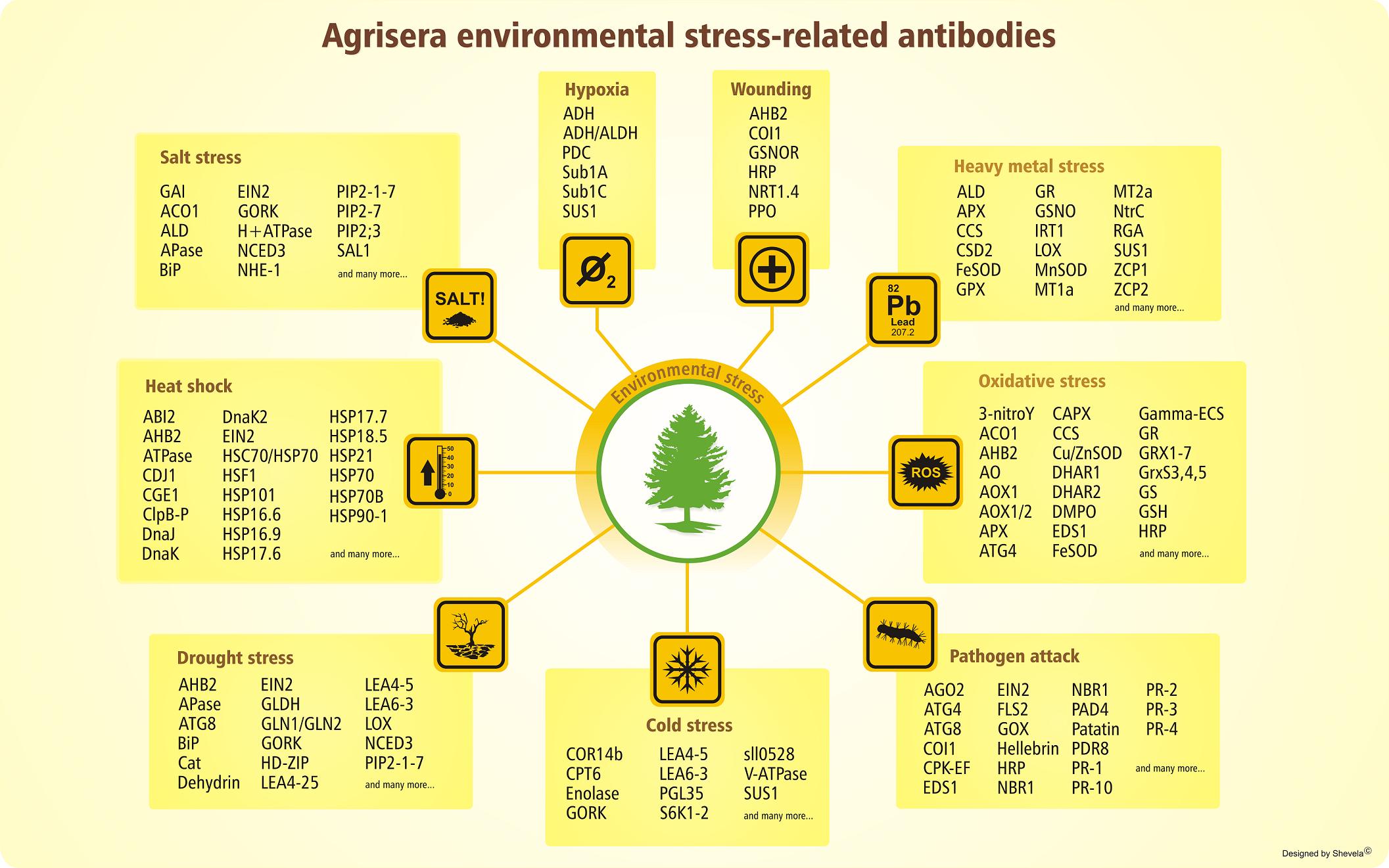
Environmental stress in Plants

Extreme temperatures, high/low light intensity, draught, flooding, salinity are, to name a few, stressors that plants will be repeatedly subjected to over the course of their life-time. As a result, due to their "sedentary life-style", plants must adapt quickly to their ever-changing environment if they want to survive. Understanding how plants sense, respond and adapt to these stressors is of primordial importance since it has recently been estimated that the temperature increase due to global warming this century will likely exceed previous predictions,[1] hence increasing environmental stress on plants not only by their intensity but also by their frequency. As plant domestication occurred under more favourable conditions than during the early evolution of land plants, crops have been selected on their productivity rather than on their resistance to abiotic stress and with the world population predicted to reach 9.7 billion by 2050, plant scientists face one of the biggest challenge of the future: increasing crop production on less area and with dwindling resources of water.[2]

How do plants respond to draught stress?
It is usually assumed that plant response to stress happens in three different phases.[3]
  1. An alarm phase, when plants detect a change in their environment and activate different mechanisms to be able to cope with the change.
  2. A resistance phase, where plants adjust their structure and function in order to withstand the stress and repair the damaged caused.
  3. If the stressor stops or lessens then the plant may recover and reach a new optimal physiological status. However, if the stress continues or is too intense, then the plant dies.
In the specific case of draught stress, plants are known to reduce photosynthesis by decreasing their leaf area as well as their photosynthesis rate, mainly by inhibiting the CO2 mechanism and by restricting the diffusion of CO2 into the leaf via stomatal closure. As a result of this and because of the low concentration of intracellular CO2, ongoing photosynthetic light reactions may cause the building-up of reduced photosynthetic electron transport components, which can react with molecular oxygen to form highly damaging reactive oxygen species (ROS).[4]

As a result, plants cope with stress by modulating key physiological processes that result in the modification of molecular and cellular processes. This plasticity is mediated by phytohormones such as abscisic acid (ABA), ethylene, cytokinin (CK), gibberellic acid (GA) and auxin as they play important roles in every step of plant development and hence will enable a plant to respond to abiotic stress. For instance, under drought stress, ABA is involved in stomatal closure whereas cytokinins are known to delay leaf senescence and death.[4]

Agrisera antibodies - Working towards a better understanding of environmental stresses in plant research

Development of crops and plants highly resilient to environmental stresses without compromising on yield is one of the many challenges that plant scientists are currently facing. To facilitate research in this area, Agrisera has developed an extensive range of plant antibodies. This includes environmental stress antibodies as well as phytohormone antibodies.


References:
1. Greater future global warming inferred from Earth’s recent energy budget, Brown PT et al, Nature 2017, 552, 45–50
2. Plant abiotic stress challenges from the changing environment; Pereira A, Front Plant Sci., 2016; 7: 1123.
3. Stress Memory and the Inevitable Effects of Drought: a physiological perspective; Fleta-Soriano E et al, Front Plant Sci, 2016,7, 143
4. Plant adaptation to drought stress, Basu S et al, Version 1. F100Res. 2016; 5: 1554.


Written by Magalie Dale
If you like my post why not connect to me on LinkedIn.

25/04/2018

Oxidative damage – The damaging effect of Reactive oxygen species ROS


Newmarket Scientific lipid peroxidation antibodies
What are ROS?
ROS, Reactive oxygen species, is a generic term to describe a range of oxygen containing radicals such as hydroxyl radical OH., superoxide anion O2-., nitric oxide NO, perhydroxyl radical HO2. and non-radical species such as hydrogen peroxide H2O2 and hypochlorous acid HOCl. They are formed as by-products during normal metabolism processes, primarily in mitochondria, but also as a cellular response to ionising radiations, pollutants, xenobiotics, cytokine and bacterial invasion.1

Oxidative stress - When the concentration of ROS becomes harmful
A certain level of ROS is important for several physiological processes such as wound healing, tissue regeneration and protection from pathogens.2,3 If the concentration of ROS increases, they will be scavenged by enzymatic oxidants (for instance such as SOD, catalase, GPx) or non-enzymatic antioxidants (e.g. vitamin C, vitamin E, transferrin, beta-carotene). However, when ROS are produced too quickly and cells are no longer able to quench them by using suitable antioxidant defences, they become harmful and can cause damage to proteins, lipid molecules of the cell membranes, carbohydrates as well as RNA and DNA. This is referred to as oxidative stress i.e. a state where ROS are overproduced and the rate of clearance via endogenous and exogeneous antioxidants is no longer sufficient to protect cells and tissues from their toxic effects. The biological consequences of oxidative stress include for instance aging, when the levels of ROS remain low but with a gradual increase or cancer when there is a rapid increase in the production of ROS resulting in a high concentration of ROS.

Oxidative stress markers
Unfortunately, ROS radicals are extremely reactive with a short half-life and consequently difficult to use as markers of oxidative stress. Nevertheless, their reactions with lipids, proteins and DNA lead to the formation of stable molecules that can be used as secondary markers of oxidative stress.

1. Protein oxidation: Oxidation of proteins can lead to fragmentation resulting in the loss of their biological activities and the formation of residues such as o-tyrosine, di-tyrosine and dibromo-tyrosine that can be used as markers of oxidative stress.

2. Lipid peroxidation: Polyunsaturated lipid molecules in cell membranes are highly susceptible to reaction with radicals via a chain reaction. This leads to the formation of lipid peroxides that can further decompose into aldehydes such as acrolein, malondialdehyde (MDA), hydroxynonenal (HNE), 4-hydroxy-2-hexenal (HNN), crotonaldehyde (CRA) and adducts such as hexanoyl-lysine (HEL) and 7-ketocholesterol (7KC). Common pathological processes linked to MDA and 4-HNE are Alzheimer’s disease, Parkinson’s disease, cancer, cardiovascular diseases and diabetes.

3. DNA damage: Oxidation of the nucleic acids can lead to the formation of 8-hydroxy 2’-deoxyguanosine, 8-OHdG. Increased levels of 8-OHdG is linked to aging as well as several pathological conditions such as cancer and diabetes and hence represents a useful marker of oxidation stress. 8-OHdG can be easily quantified using ELISA kits from urine or complex samples such as plasma, cell lysates and tissues.

Tools for oxidative stress research

StressMarq has developed an extensive range of products to study oxidative stress. These products are currently available in the UK and Ireland through Newmarket Scientific and include:

References:
1. Ray PD et al, Cell Signal. 2012; 24 (5):981-990
2. Onodera Y et al, FEBS Open Bio, 2015; 5: 492–501.
3. Di Meo et al, Oxid Med Cell Longev. 2016; 2016: 7909186.

Written by Magalie Dale
If you like my post why not connect to me on LinkedIn.

26/03/2018

Stopping Index Hopping in NGS

Increased mis-assignment of indexes has been shown to occur on Illumina® sequencing instruments that feature a patterned flow cell and exclusion amplification technology. This mis-assignment of indexes, also known as index hopping, index switching or spread of signal, is acknowledged by Illumina on their website.

Illumina® Website (March 2018):
"Index hopping or index switching is a known phenomenon that has impacted NGS technologies from the time sample multiplexing was developed. It causes a specific type of misassignment that results in the incorrect assignment of libraries from the expected index to a different index (in the multiplexed pool).

Index hopping can be seen at slightly elevated levels on instruments using patterned flow cells with exclusion amplification chemistry versus those that do not use patterned flow cells. Libraries with higher levels of free adapters will see higher levels of index hopping."


The phenomenon has been described in a preprint paper (by Rahul Sinha et al.) that suggests the issue might occur during the act of sequencing itself, with free oligos in the pool somehow associating with the fragments being sequenced and causing the fragment to be identified with the index of the free oligo as opposed to it’s true original sequence.

Any single index system may be subject to this phenomenon, but the NEXTFLEX® unique dual index barcodes have been designed to specifically mitigate the index hopping phenomenon. With unique dual index barcodes, if a free oligo associates with a fragment causing a "change" in the index of the read, the other index associated with the fragment will show the read is incorrect and prevent it from being associated with a given sample.

It is important to note however that not all dual index systems would address this problem, as standard dual indexes using a combinatorial approach (using the same first index across sets of a second index) would still lead to the incorrect allocation of some reads as shown below.

In the standard dual indexing example below (combinatorial dual indexing), the fragment was originally indexed with the blue and purple indexes, but it now being identified as indexed with the gold and purple indexes. However because there is also a sample present using the gold and purple index combination, this read is now going to be incorrectly included in that other sample.


However if this same situation occurs with the UNIQUE dual indexes, the resultant contaminant read will not be added to another sample present. The incorrect read will still occur, but this time it isn’t reassigned to another sample present, as no sample contains that combination of indexes. Both indexes are unique so the issue is avoided.

Index mis-assignment can lead to increased false positive rates, which are especially detrimental to sensitive applications.

Multiplexing with NEXTFLEX® unique dual index barcodes significantly increases processing capacity while reducing costs by allowing the user to pool multiple libraries in a single flow cell lane, whilst providing unprecedented data security in sequencing applications.


Written by Rick Bhatt
If you like my post why not connect to me on LinkedIn.

26/02/2018

NS Reagents Antibodies - Now Available

NS Reagents is the new Newmarket Scientific range of antibodies focussing on Neuroscience and DNA Damage Repair & Methylation, but these are just the areas we are starting with and we have antibodies for other research areas in development, which we will release over the coming months.

Anti-TDP43 (Clone DB9)
Anti-TDP43 (Clone DB9) TAR DNA-binding protein 43 (TDP-43) has been shown to bind both DNA and RNA and has multiple functions in transcriptional repression, pre-mRNA splicing and translational regulation. It belongs to the hnRNP protein family and is highly expressed in the pancreas, placenta, lung, genital tract and spleen. Characterisation of transcriptome-wide binding sites revealed that thousands of RNAs are bound by TDP-43 in neurons.

TDP-43 regulates alternate splicing of the CFTR gene. The resulting aberrant splicing is associated with pathological features typical of cystic fibrosis. Mutations in TDP-43 have been associated with amyotrophic lateral sclerosis, frontotemporal dementia, Parkinson's disease and Alzheimer's disease.

Applications: ELISA ¦ IHC ¦ WB   Host: Mouse   Reactivity: Human


Anti-DNA damage-binding protein 1
DNA damage-binding protein 1 (DDB1) is the large subunit (p127) of the heterodimeric DNA damage-binding (DDB) complex. DDB1 also functions as a core component of the cullin 4 (CUL4) ubiquitin E3 ligase complex, facilitating the binding of substrates to this complex and the ubiquitination of proteins. These factors (ubiquitin ligase substrates) regulate numerous essential processes in the cell including DNA repair (DDB2), DNA replication, chromatin remodelling (Cdt2) and more.

Applications: WB      Host: Rabbit      Reactivity: Human


Details of the full antibody range is available on our website at https://www.newmarketscientific.com/nsreagents

15/01/2018

True optimisation of antibodies for immunohistochemistry

Immunohistochemistry (IHC) is often considered something of a "black art". As well as requiring a high degree of knowledge of the techniques involved, it generally requires an element of trial and error to have a given antibody work well with a given tissue and frequently necessitates the use of antibodies and techniques tailored to each individual project or diagnostic application.

This means, there is no one specific protocol for IHC that can be used regularly.

Antibody optimisation for IHC involves a range of tests in order to find an antibodies optimal staining conditions. Each antigen has a preferred method of antigen retrieval such as Heat Induced Epitope Retrieval (HIER) using acidic Citrate or TRIS-EDTA base buffers, as well as an enzymatic retrieval process. However the majority of antigens need an alkaline pre-treatment method for optimal staining. Additionally each antibody has an optimal concentration when used, depending on the affinity of paratope and epitope, as well as the expression level of the antigen.

Antibodies optimised with tissues that express high levels of antigen may prove inadequate when staining tissues with low antigen expression. For this reason it is necessary to optimise each antibody for a variety of tissue types.

Additionally from a clinical perspective, antibodies must be specific with high affinity towards their epitopes, whilst remaining flexible to use and offering good LOT consistency.

This is the basis of our Optibodies range with each antibody optimised for each use.

Optibodies are a range of carefully tested, top quality antibodies for Immunohistochemistry that has been carefully optimised and fine-tuned for both research and clinical IHC using NordiQC recommended control tissues and criteria.

Optibodies (TM) from Nordic Biosite - Optimal Antibodies for Optimal Immunohistochemistry

Details of the full Optibodies antibody range are available here.


EpCAM

Her2

PDL1

SYP

27/11/2017

HSP70

HSP70s are monomeric proteins that reside in the cytosol of prokaryotes and the cytosol, nuclei, ER, mitochondria and chloroplasts of eukaryotes 8. In addition to their intracellular location, HSP70s have been found in the plasma membrane of malignantly transformed cells, on virally / bacterially infected cells and in the extracellular space. Extracellular Hsp70 exists in a free soluble form, complexed to antigenic peptides, or in exosomes 12,13,14,15. Noteworthy, Hsp70-1 as the most prominent member of the HSP70 family can be detected in the plasma membrane of a large proportion of different tumor entities, but not in the plasma membrane of normal cells/tissues 16,17,18. Several observations have led to the hypothesis that in tumor cells Hsp70-1 is an integral membrane protein associated with certain membrane lipid components 15, 16.

Fig 1: Ribbon and tube representation of the tertiary Hsp70-1 structure in the presence of ADP.

Heat shock proteins (HSPs) were originally described in the early 1960s by the pioneering work of Ferruccio Ritossa on the fruit fly Drosophila melanogaster 5,6,7. Expression of HSPs was found as being induced after exposure to different kinds of stress such as heat shock and could be demonstrated subsequently in any cellular organism 8. Nevertheless, other stress conditions, including heavy metals, hypoxia, nutrient deprivation and irradiation as well as oxidative and toxic stress, infections and exposure to inflammatory cytokines are also able to induce HSP expression 8, 9. Members of the HSP70 family were identified for the first time as being upregulated in bacteria in response to cellular stress 10. The painstaking analysis of the limited number of proteins firstly identified by heat shock induction in D. melanogaster and in E. coli led to the finding that DnaK, DnaJ and GrpE were also members of the heat shock class of proteins. In 1984, Bardwell and Craig demonstrated that the E. coli DnaK and the Drosophila 70 kDa heat shock proteins were highly conserved at the sequence level 11. Moreover, hybridization between the DNA of the archaebacterium Methanosarcina barkeri and the HSP70 genes of D. melanogaster, Saccharomyces cerevisiae, and E. coli has been detected, suggesting the existence of Hsp70-related genes in the three “primary kingdoms”: eukaryotes, eubacteria, and archaebacteria11.

The HSP70 Family

The HSP70 family represents the most conserved and best characterized group of HSPs comprising polypeptides whose molecular weights range from 66 – 78 kDa and that are encoded by a multigene family encompassing up to 17 genes and 30 pseudogenes in humans 19. Functional genes encoding HSP70 proteins map to human chromosomes 6, 14, 21, and at least one other chromosome 20. The most studied genes are HSPA1A and HSPA1B encoding proteins that only differ by two amino acids and are believed as being completely interchangeable proteins. The genes are clustered in the major histocompatibility complex class III region on chromosome 6p21.3.

The HSP70 family constitutes one of the most conserved protein families in evolution. Their members are present in all organisms and subcellular compartments and can be found from archaebacteria and plants to humans. In archaea and eubacteria Hsp70 is referred to as DnaK. In yeasts they are called Ssa, in mammals including humans they are referred to as HspA.

More information is available on the HSP70 website
and antibodies and related products are available by searching on the Newmarket Scientific website.

References:
1. Gribaldo,S. et al. Discontinuous occurrence of the hsp70 (dnaK) gene among Archaea and sequence features of HSP70 suggest a novel outlook on phylogenies inferred from this protein. J. Bacteriol.181, 434-443 (1999). [PubMed]

2. Sharma,D. & Masison,D.C. Hsp70 structure, function, regulation and influence on yeast prions. Protein Pept. Lett.16, 571-581 (2009). [PubMed]

3. Sharma,D. et al. Function of SSA subfamily of Hsp70 within and across species varies widely in complementing Saccharomyces cerevisiae cell growth and prion propagation. PLoS. ONE.4, e6644 (2009). [PubMed]

4. Kampinga,H.H. & Craig,E.A. The HSP70 chaperone machinery: J proteins as drivers of functional specificity. Nat. Rev. Mol. Cell Biol.11, 579-592 (2010). [PubMed]

5. Ritossa,F. Experimental activation of specific loci in polytene chromosomes of Drosophila. Exp. Cell Res.35, 601-607 (1963). DOI: 10.1016/0014-4827(64)90147-8

6. Ritossa,F. New puffs induced by temperature shock, DNP and salicilate in salivary chromosomes of D. melanogaster. Drosophila Information Service37, 122-123 (1963). [Drosophila Information Service]

7. Ritossa,F. A new puffing pattern induced by temperature shock and DNP in Drosophila. Experientia18, 571-573 (1962). DOI: 10.1007/BF02172188

8. Lindquist,S. & Craig,E.A. The heat-shock proteins. Annu. Rev. Genet.22, 631-677 (1988). [PubMed]

9. Jäättelä,M. Heat shock proteins as cellular lifeguards. Ann. Med.31, 261-271 (1999). [PubMed]

10. Craig,E.A. & Gross,C.A. Is hsp70 the cellular thermometer? Trends Biochem. Sci.16, 135-140 (1991). [PubMed]

11. Bardwell,J.C. & Craig,E.A. Major heat shock gene of Drosophila and the Escherichia coli heat-inducible dnaK gene are homologous. Proc. Natl. Acad. Sci. U. S. A81, 848-852 (1984). [PubMed]

12. Bausero,M.A., Gastpar,R., Multhoff,G., & Asea,A. Alternative mechanism by which IFN-gamma enhances tumor recognition: active release of heat shock protein 72. J. Immunol.175, 2900-2912 (2005). [PubMed]

13. Gastpar,R. et al. Heat shock protein 70 surface-positive tumor exosomes stimulate migratory and cytolytic activity of natural killer cells. Cancer Res.65, 5238-5247 (2005). [PubMed]

14. Lancaster,G.I. & Febbraio,M.A. Exosome-dependent trafficking of HSP70: a novel secretory pathway for cellular stress proteins. J. Biol. Chem.280, 23349-23355 (2005). [PubMed]

15. Vega,V.L. et al. Hsp70 translocates into the plasma membrane after stress and is released into the extracellular environment in a membrane-associated form that activates macrophages. J. Immunol.180, 4299-4307 (2008). [PubMed]

16. Gehrmann,M. et al. Tumor-specific Hsp70 plasma membrane localization is enabled by the glycosphingolipid Gb3. PLoS. ONE.3, e1925 (2008). [PubMed]

17. Schilling,D. et al. Binding of heat shock protein 70 to extracellular phosphatidylserine promotes killing of normoxic and hypoxic tumor cells. FASEB J.23, 2467-2477 (2009). [PubMed]

18. Stangl,S. et al. Targeting membrane heat-shock protein 70 (Hsp70) on tumors by cmHsp70.1 antibody. Proc. Natl. Acad. Sci. U. S. A108, 733-738 (2011). [PubMed]

19. Brocchieri,L., Conway de,M.E., & Macario,A.J. hsp70 genes in the human genome: Conservation and differentiation patterns predict a wide array of overlapping and specialized functions. BMC. Evol. Biol.8, 19 (2008). [PubMed]

20. Harrison,G.S. et al. Chromosomal location of human genes encoding major heat-shock protein HSP70. Somat. Cell Mol. Genet.13, 119-130 (1987). [PubMed]


25/10/2017

A unique kit for the detection of Dibromo-tyrosine

Dibromo-tyrosine is produced by the oxidative bromination of tyrosine residues. This reaction occurs via eosinophil peroxidase (EPO), an enzyme released by activated eosinophils. Upon activation of eosinophils, a respiratory burst occurs releasing elevated levels of O2 and H202. In the oxidation of tyrosine, EPO utilises H202 to catalyse the peroxidation of physiological levels of bromine found within plasma to generate the brominating reagent hypobromous acid (HOBr) (Ref: 1-5)


Figure 1. Bromination of tyrosine (Ref: 8)

Eosinophils play an immunomodulatory role through their recruitment to host sites of parasitic invasion. EPO levels also contribute to diseases such as asthma, cancers and allergic disorders where cellular activation is found to occur at pathological sites (Ref: 6-10)

Brominated products such as 3,5-dibromo-tyrosine serve as biological markers for in vivo eosinophil-mediated tissue damage which allows for understanding the overall roll oxidative stress has on pathways implicated in diseased states within organisms (Ref: 4) .


About This Assay

This unique kit is part of the StressMarq range. It is a is a competitive ELISA assay that can be used for the quantification of 3,5-dibromo-tyrosine in urine, plasma, and other sample matrices. The assay utilises a dibromo-tyrosine-coated plate and an biotin-conjugated antibody for detection which provides an assay range of 0.078 - 5 μg/mL, with a sensitivity of 0.04 μg/mL. Additional kit highlights are quick incubation times, stable reagents, and an easy to use protocol.

It is important to note that the dibromo-tyrosine antibody used in this assay recognises both free dibromo-tyrosine and brominated residues within a protein. Since complex samples such as plasma, are comprised of mixtures of protein fragments and free 3,5-dibromo-tyrosine, concentrations of 3,5-dibromo-tyrosine reported by ELISA methodology may not coincide with literature values where the free residue is typically measured. This should be kept in mind when analysing and interpreting experimental results.


Assay Overview


Figure 2. Schematic of the dibromo-tyrosine competitive ELISA

    Further details for this kit are available here:
    Dibromo-tyrosine ELISA Kit Details and Pricing

    References:
    1. MacPherson, J.C., Comhair, S. A. A., Erzurum, S.C., et al. Eosinophils are a major source of nitric oxide-derived oxidants in severe asthma: characterization of pathways available to eosinophils for generating reactive nitrogen species. J. Immun. 166, 5763-577 (2001).
    2. Mayeno, A. N., Curran, A. J., Roberts, R. L., et al. Eosinophils Preferentially Use Bromide to Generate Halogenating Agents. J. Biol. Chem. 264, 5660-5668 (1989).
    3. Babior, B. M. Oxygen-dependent microbial killing by phagocytes. N. Engl. J. Med. 298, 659-668 (1978).
    4. Wu W., Chen, Y., d’Avignon, A. et al. 3-Bromotyrosine and 3,5-dibromotyrosine are major products of protein oxidation by eosinophil peroxidase: potential markers for eosinophil-dependent tissue injury in vivo. Biochem. 38, 3538-3548 (1999)
    5. Kambayashi, Y., Ogino, K., Takemoto, K. et al. Preparation and characterization of a polyclonal antibody against brominated protein. J. Clin. Biochem. Nutr. 44, 95-103 2009
    6. Wang J., Slungaard A. Role of eosinophil peroxidase in host defense and disease pathology. Arch. Biochem. Biophys. 445, 256–260 (2006).
    7. Kazura, J. W., Fanning, M. M., Blumer, J. L. Mahmoud, A. A. Role of cellgenerated hydrogen peroxide in granulocyte-mediated killing of schistosomula of Schistosoma mansoni in vitro. J. Clin. Invest. 67, 93 (1981).
    8. Klebanoff, S. J., Locksley, R. M., Jong, E. C., Rosen, H. Oxidative response of phagocytes to parasite invasion. CIBA Found. Symp. 99: 92 (1983)
    9. Gleich, G. J., Ottesen, E. A., Leiferman, K. M., Ackerman, S. J. Eosinophils and human disease. Int. Arc. Allergy Appl. Immunol. 88: 59 (1989).
    10. Wardlaw, A. J., Eosinophils in the 1990s: new perspectives on their role in health and disease. Postgrad. Med. J. 70: 536 (1994).